Surgical Vignettes: Callot’s Triangle
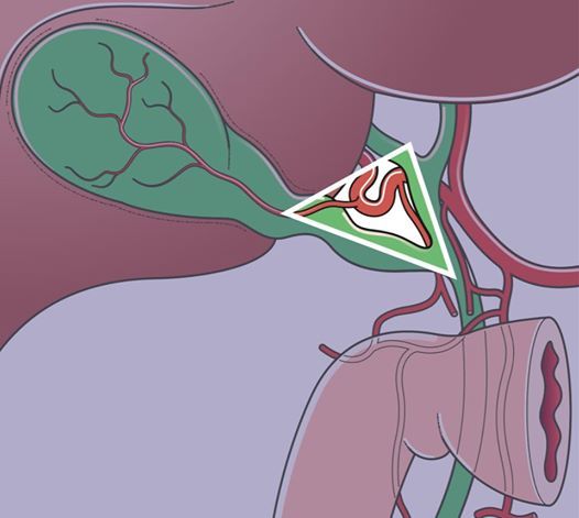
Ref: Bailey and Love 26th ed (http://amzn.to/1OdJGT3)Calot’s triangle or the hepatobiliary triangle is the space bordered by the cystic duct inferiorly, the common hepatic artery medially and the superior border of the cystic artery. This was described in 1891 by Jean-François Calot. It is an important surgical landmark and should be identified by surgeons performing a cholecystectomy to avoid damage to extrahepatic billiary system.The most dangerous anomalies of callot’s triangle are where the hepatic artery takes a tortuous course on the front of the origin of the cystic duct, or the right hepatic artery is tortuous and the cystic artery short. The tortuosity is known as the ‘caterpillar turn’ or ‘Moynihan’s hump’ (Figure attached). This variation is the cause of many problems during a difficult cholecystectomy with inflammation in the region of the cystic duct.