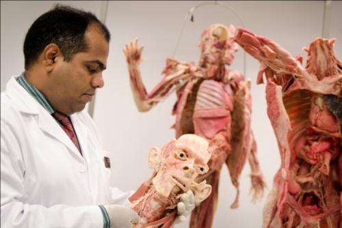
Nanyang Technological University’s (NTU) new medical school will be pioneering the use of plastinated bodies for medical education in Singapore. Plastinated bodies are human bodies which people have pledged towards the learning and advancement of science upon their death and are preserved through plastination. These human bodies are preserved by replacing the fat and water in body tissues with plastic. The human bodies and body parts, known as specimens, will be used in anatomy classes taught at the Lee Kong Chian School of Medicine (LKCMedicine), and will be ready for the first batch of 54 medical students this August. The new medical school, jointly set up by NTU and Imperial College London, had ordered the human bodies and specimens from Germany, which were prepared to the school’s specifications.
There are two whole bodies and various body parts such as hearts, lungs, brains, limbs and torsos. With the use of plastinated bodies and specimens, traditionally preserved cadavers which are in short supply in Singapore, will no longer be needed by LKCMedicine. There are many distinct advantages of using plastinated specimens over traditional cadavers, making it an excellent teaching tool. Top medical schools around the world that use plastinated specimens include Warwick University in UK and, New York University in the United States. These advantages include excellent quality and exceptional durability of the specimens; “high-resolution” intricate details of arteries, veins, muscle and nerves and greater flexibility of use both inside and outside of the classroom, including the ability to interface with computer software. Asst Professor Dinesh Kumar Srinivasan, the Lead for Anatomy Teaching at LKCMedicine, said the plastinated human specimens are excellent tools for teaching gross anatomy and neuroanatomy, as it is durable, safe and non-toxic.
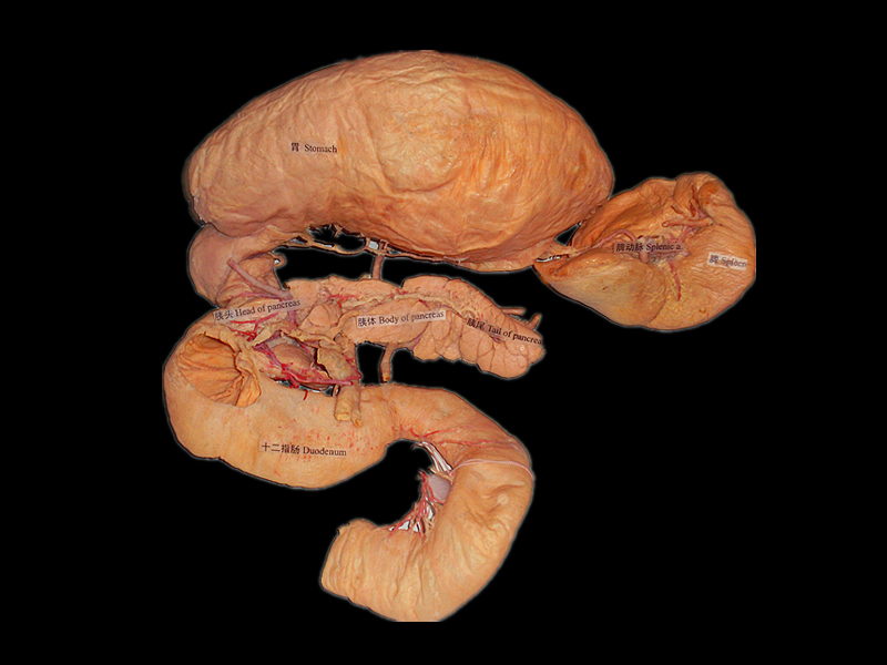
“These highly detailed, plastinated human specimens are very durable and can be repeatedly handled by students without deterioration and it can be stored just like any inert object,” Prof Srinivasan said. “They may be used in a much broader range of educational settings, since we no longer have to take extra steps to re-preserve the body in embalming chemicals, as we would need to for traditional cadavers each time they are used in class. “In addition, plastinated human specimens can be used to demonstrate difficult structures and dissection areas in ‘high-definition’, such as the blood vessels in the brain or the nerves in the spine. Such intricate details, essential in learning anatomy, will start degrading in traditionally preserved cadavers after repeated use.” Importance of anatomy in medicine NTU’s investment in a collection of high-quality, plastinated real human bodies and body parts underlines its commitment to the importance of anatomy education as the basic foundation of medical sciences.
The knowledge of structure of the human body from the naked eye appearance (gross anatomy) down to the molecular level is fundamental to understanding its function, and how both structure and function are modified by disease processes. Students at LKCMedicine will be learning anatomy in their first and second year to build a strong foundation for future medical knowledge. In their third and fourth year, anatomy education is important due to the students’ clinical exposure in hospitals, where the relevance of anatomy to clinical practice will be continuously emphasised, Prof Srinivasan said. “The anatomy lab experience will be very different for a third-year student as compared to a first-year medical student. First-year students will be mostly focused on identifying structures from their textbook, whereas third-year student will be interested in how the anatomy has clinical value and how it relates to real life patients,” Prof Srinivasan added.
First class anatomy education At LKCMedicine, anatomy will be taught in a clinically orientated manner so as to prepare students for their clinical studies. All anatomy practical lessons will be conducted in a small group environment, with hands-on practice emphasising clinical relevance to medical practice. In addition to plastinated human bodies, students will also learn from microscopic slides to understand the microscopic anatomy of cells and tissues of humans. From these histology slides, students will be able to see at a microscopic level how healthy and diseased cells look like, which aids in their understanding of cells and how they are affected by various illnesses. Anatomy classes will be also supplemented by the use of the Anatomage Table, which displays life-sized 3D images of full body anatomy using computed tomography (CT) and magnetic resonance imaging (MRI) scans, allowing students to view the body’s internal structures. LKCMedicine is the first medical school in Singapore to adopt the Anatomage Table which arrived earlier in March this year. The combination of the plastinated human specimens, Anatomage Table and histology slides will enable students to learn about the entire human body from the outside to inside, and down to the cellular level, said Prof Srinivasan.
“With the new plastinated bodies and the interactive Anatomage Table, we believe this will help inspire the students to keep up lifelong learning. Such cutting-edge teaching tools are one of the benefits a new medical school has, where we can plan for world-class facilities and a clinically-relavent curriculum.” “The teaching of anatomy will involve input, ideas, and perspectives from surgeons and radiologists, who are in clinical practice,” he added. In future, students will also have opportunities to learn anatomical structures with their function from experienced surgeons and radiologists.”