Q-1. Abnormality shown in the following picture?
a) Galleazzi
b) Monteggia
c) Smith
d) Colles
Answer: Galleazzi
Explanation:
Galleazzi fracture or Reverse Monteggia fracture:
Galeazzi fracture-dislocations consist of fracture of the distal part of the radius with dislocation of distal radio-ulnar joint and an intact ulna.
Important point:
Monteggia fracture-dislocations comprise of a fracture of the ulna shaft and dislocation of the radial head.
Colles fractures are very common extra-articular fractures of the distal radius that occur as the result of a fall onto an outstretched hand. They consist of a fracture of the distal radial metaphyseal region with dorsal angulation and impaction, but without involvement of the articular surface.
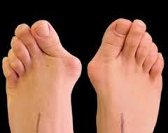
Q-3. Identify the test being performed by a patient in the image below?
a) Hallux valgus
b) Hallux varus
c) Rheumatoid nodule
d) Sub-cutaneous nodule
Answer: Hallux valgus
Explanation:
Hallux valgus is considered to be a medial deviation of the first metatarsal and lateral deviation and/or rotation of the hallux, with or without medial soft-tissue enlargement of the first metatarsal head.
The Keller procedure is an excision arthroplasty of the 1st MTP joint, which can be used for both Hallux rigidus and valgus.
Q-2. AP & Lat. view of wrist is given. Diagnosis is
a) Galleazzi
b) Monteggia
c) Smith
d) Colles
Answer: Galleazzi
Explanation:
Galleazzi fracture or Reverse Monteggia fracture:
Galeazzi fracture-dislocations consist of fracture of the distal part of the radius with dislocation of distal radio-ulnar joint and an intact ulna.
Important point:
Monteggia fracture-dislocations comprise of a fracture of the ulna shaft and dislocation of the radial head.
Colles fractures are very common extra-articular fractures of the distal radius that occur as the result of a fall onto an outstretched hand. They consist of a fracture of the distal radial metaphyseal region with dorsal angulation and impaction, but without involvement of the articular surface.
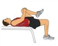
Q-3. Identify the test being performed by a patient in the image below?

Or
Hip flexion test is
a) Trendelenburg
b) Thomas test
c) Smith
d) Narath
Answer: Thomas test
Explanation:
Thomas test is used to check for hip flexion contractures.
The hip flexors:
Psoas major
Iliacus muscle
Rectus femoris
Tensor fasciae latae
Q-4. Identify the deformity in the image below?
a) Humerus lateral condylar fracture
b) Medial condylar fracture of humerus
c) Fracture shaft humerus
d) Medial condylar fracture of humerus
Answer: Humerus lateral condylar fracture
Explanation:
Humerus lateral condylar fracture:
It is Salter and Harris type-IV epiphyseal injury.
It is common and occurs due to the pull of the common extensor muscles which take origin from the epicondyle.
The fragment is rotated outwards along its vertical and horizontal axes; sometimes even as much as 90 degree.