
Case Presentation:
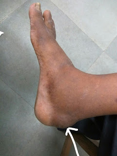
A 60 year old male patient came with complaint of swelling and pain over posterior aspect of ankle. He thinks that it was caused by a blunt injury over the same site about 2 years back after which he developed the swelling which gradually grew in size to attain the current size and has been having chronic pain while walking ever since. The swelling is about 3x3cm in size, not warm, tender, firm and not freely mobile. Plain of the swelling is subcutaneous.On Xray there was no bony prominence corresponding to this swelling.
Case discussion on Subcutaneous Calcaneal Bursitis
Anatomy of Ankle Bursae
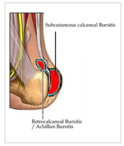
Pain at the posterior aspect of ankle is most commonly due to pathology at the posterior calcaneus, the Achilles (calcaneal) tendon, or the associated bursae. Following are the associated bursae [1]:
Subtendinous calcaneal bursa: Also called the retrocalcaneal bursa, situated anterior (deep) to the Achilles tendon, is located between the Achilles tendon and the calcaneus
Subcutaneous calcaneal bursa: Also called the Achilles bursa, it is found posterior (superficial) to the Achilles tendon, lying between the skin and the posterior aspect of the distal Achilles tendon
Case discussion on Subcutaneous Calcaneal Bursitis
Pathophysiology of Ankle Bursitis
Inflammation of the calcaneal bursae is most commonly caused by repetitive overuse and cumulative trauma, as seen in runners wearing tight-fitting shoes. Such bursitis may also be associated with conditions such as gout, rheumatoid arthritis, and seronegative spondyloarthropathies.
This is not same as Haglund’s deformity. Haglund deformity, also known as a pump bump or Bauer bump or Mulholland deformity, is defined as bony enlargement formed at posterosuperior aspect of calcaneum. This deformity leads to retrocalcaneal bursitis [2].
References:
[1] Kachlik D, Baca V, Cepelik M, et al. Clinical anatomy of the retrocalcaneal bursa. Surg Radiol Anat. 2008 Mar 11
[2] Haglund deformity | Radiology Reference Article | Radiopaedia.org